


Introduction
Ectopic pregnancy occurs in 0.6% to 2.1% of all pregnancies [1] and accounts for up to 5% of all maternal deaths in high-resource countries [1,2]. It is also the main cause of mortality during the first trimester of pregnancy [1,3,4,5,6]. Laparoscopic tube-preserving surgery including salpingotomy, salpingostomy, segmental tubal resection and reanastomosis, and fimbrial milking is a well-established treatment for ectopic tubal pregnancy patients who desire to retain their reproductive potential [2,4,6,7,8,9]. According to a recent study assessing patientsŌĆÖ preferred surgical method (tube-preserving surgery vs. salpingectomy) [10], most women (88%) preferred tube-preserving surgery despite the resultant risk of persistent trophoblast and, possibly, a repeat ectopic pregnancy in the operated tube [5].
However, laparoscopic tube-preserving surgery for tubal pregnancy has yet to be widely adopted. It is therefore necessary to gather data on these procedures in order to make more information available to women with tubal pregnancy. The aim of this study was to present our experience with laparoscopic tube-preserving surgery for tubal pregnancy and to evaluate its feasibility and efficacy.
Materials and methods
1. Study patients
After Institutional Review Board approval, this study was conducted prospectively at two institutions (CHA Gangnam Medical Center between February 2012 and May 2014, Kangbuk Samsung Hospital between June 2014 and March 2016). Patients who were diagnosed with an ectopic tubal pregnancy and selected laparoscopic surgery were approached for study enrollment. The diagnosis of tubal pregnancy was made by using a non-laparoscopic approach using transvaginal sonography, ╬▓-human chorionic gonadotropin (╬▓-hCG) levels, or clinical examination [3]. Preoperative informed consent was obtained from all patients after providing explanations of the possible risks and complications of tube-preserving surgery such as persistent trophoblast and a repeat ectopic pregnancy in the operated tube.
The inclusion criteria were as follows: a visible ectopic tubal mass on transvaginal sonography; a willingness to preserve fertility; a desire to maintain optimal tubal patency for future fertility; at least 18 years of age; an appropriate medical status for laparoscopic surgery (American Society of Anesthesiologists Physical Status classification 1 or 2); and consent to surgical treatment and follow-up. The exclusion criteria were as follows: no desire for future pregnancy; an interstitial or heterotopic pregnancy; pregnancy by in vitro fertilization; pregnancy in a solitary tube; or a contralateral tubal occlusion or hydrosalpinx, either documented earlier at hysterosalpingography or laparoscopy or detected during surgery for the index ectopic pregnancy.
2. Surgical procedures
A single surgeon (ST) performed all surgical procedures including salpingotomy, salpingostomy, segmental resection and reanastomosis, and fimbrial milking. All patients received the same standard surgical preparation, which included the administration of prophylactic antibiotic therapy 30 minutes before the procedure. After the induction of general anesthesia via endotracheal intubation, patients were placed in the deep Trendelenburg position. In all cases, a 12-mm trocar was inserted in the umbilicus and three 5-mm trocars were inserted in the lower abdomen. The abdominal cavity was then carefully examined, and a suction device was used to evacuate the hemoperitoneum, if present.
1) Salpingotomy or salpingostomy
For salpingotomy or salpingostomy, a dilute solution of vasopressin (Hanlim Pharm, Seoul, Korea; 2.5 U vasopressin in 10 mL saline solution) was injected through a 23-gauze spinal needle into the mesosalpinx around the affected tube to reduce blood loss. After immobilizing the involved tube with an atraumatic grasper, a linear incision was made to allow for the removal of the ectopic mass in its entirety. A fine-tip needle cautery was used to make the incision in the superior aspect of the fallopian tube just above its largest diameter, correlating with the location of the ectopic pregnancy. The ectopic mass was then removed using a combination of hydrodissection and traction using atraumatic forceps. Aspiration and compression lateral to the incision site were applied to facilitate the removal of the products of conception, if necessary. In cases of a ruptured tubal pregnancy, the product of conception was evacuated from the ruptured site without a separate incision. After the entirety of the pregnancy was removed, the placental bed was carefully evaluated. In the event of bleeding or oozing of blood from inside the lumen or tubal wall, bleeding sites were covered with a hemostatic sealant (FloSeal, Baxter Healthcare Corporation, Deerfield, IL, USA) under direct vision with a laparoscopic applicator, followed by a 2-minute wait for the FloSeal to act (Supplemental Video available at http://youtu.be/f5Yipi8DuEI). Bleeding sites were then reexamined by irrigation. For salpingotomy, once hemostasis was achieved, the tube was closed in a single layer by two or three interrupted stitches using 6-0 PDS (Ethicon, Somerville, NJ, USA). For salpingostomy, the tubal lumen was not closed and was allowed to heal by secondary intention (Supplemental Video available at http://youtu.be/lrbLT7-2ySI).
2) Segmental resection and reanastomosis
Segmental resection and reanastomosis was performed in cases of (1) unruptured ectopic pregnancy in the isthmus of the tube, (2) uncontrolled bleeding from the tubal implantation site during salpingo(s)tomy, or (3) excessive use of cautery on the placental bed to obtain hemostasis. The procedure begins with the subserosal injection of diluted vasopressin into both the proximal and distal ends and into the mesosalpinx to facilitate hemostasis and subsequent dissection of both ends. The ectopic mass and its surrounding tube are then sharply resected with laparoscopic scissors. Next, the mesosalpinx is approximated using a series of 5-0 PDS stitches to bridge the gap between the two ends of the fallopian tube and to prevent tension at the anastomosis site. In cases of a major discrepancy in size between the proximal and distal anastomosis sites, an intratubal splint with 2-0 monofilament nylon is placed to facilitate the suturing of the two ends. The mucosal and muscular layers of the tubal segments are sutured with four interrupted 6-0 PDS sutures. The first suture is placed at 6 oŌĆÖclock in such a way that the knot lies on the outside of the lumen and tied using intracorporeal knot tying techniques. The second and third sutures are placed at 3 oŌĆÖclock and 9 oŌĆÖclock, respectively, but not tied. The fourth stitch is then placed at 12 oŌĆÖclock, and the 3 and 9 oŌĆÖclock stitches are tied. Attention to proper suturing will avoid misalignment or rotation of the distal tubal segment along its longitudinal axis. The serosa is then closed separately with 5-0 PDS sutures. The immediate success of the procedure is evaluated using chromopertubation to document tubal patency.
3) Fimbrial milking
When the product of conception was located at the fimbrial end or very close to the fimbria, it was removed by grasping the tubal segment and stepwise milking the gestational product out of the tubeŌĆÖs fimbriae. The product of conception was gently pushed until extrusion. The stepwise movements began in the proximal part of the tube and the product was gently pushed into the abdominal cavity.
The surgical specimens were then removed using a laparoscopic bag (LapBag, Sejong Medical, Paju, Korea). Subsequently, the contralateral fallopian tube was closely inspected to exclude the possibility of unexpected tubal lesions (i.e., hydrosalpinx, severe peritubal adhesions, malformations, or other pathologies) and copiously irrigated with 3,000 to 4,000 mL of normal saline if a hemoperitoneum was present. To prevent the development of persistent trophoblast [11,12,13], a 50-mg/m2 intramuscular injection of methotrexate was routinely administrated within a first 24-hour after surgery, except patients who received methotrexate therapy within a week before surgery or patients who switched from tube-preserving surgery to salpingectomy.
Patients were discharged on postoperative day 2 or 3, and their serum ╬▓-hCG levels were monitored on a weekly basis until they were lower than 5 mIU/mL to identify persistent trophoblast. The ╬▓-hCG resolution time was defined as the period from the date of the surgery to the date that a serum ╬▓-hCG level less than 5 mIU/mL was achieved. Patients were diagnosed with persistent trophoblast when the postoperative serum ╬▓-hCG concentration increased again or did not decrease for at least 1 week [14]. To assess patency of the affected tube after surgery, patients were offered a hysterosalpingogram 3 months after achieving a ╬▓-hCG level less than 5 mIU/mL. Tubal patency tests were assessed by a gynecologic radiologist unaware of the patientŌĆÖs treatment.
3. Outcome measures
The primary outcome of this study was the success of tube-preserving surgery without the need for salpingectomy. Secondary outcomes included (1) postoperative complications related to tube-preserving surgery such as persistent trophoblast or re-operation; and (2) tubal patency assessed using a hysterosalpingogram.
Statistical analysis was carried out using IBM SPSS ver. 20.0 (IBM Corp., Armonk, NY, USA). After an assessment of normal distribution, data were presented as the mean┬▒standard deviation or the median (range) for quantitative variables. Qualitative variables were presented as the frequency (percentage).
Results
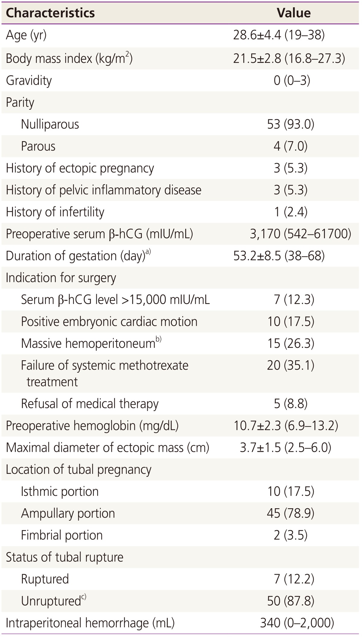
During the 4-year study period, 57 consecutive patients with tubal pregnancy were enrolled in this prospective cohort study. The demographic characteristics of the study population are displayed in Table 1. At the time of surgery, the mean patient age was 28.6┬▒4.4 years (range, 19 to 38 years) and the mean body mass index was 21.5┬▒2.8 kg/m2 (range, 16.8 to 27.3 kg/m2). All but four patients were nulliparous. Three patients (5.3%) had a history of an ectopic pregnancy that was managed with systemic methotrexate treatment. Three patients (5.3%) had a history of pelvic inflammatory disease that was managed with antibiotics. The median preoperative serum ╬▓-hCG level was 31,700 mIU/mL (range, 542 to 61,700 mIU/mL) and the mean duration of gestation was 53.2┬▒8.5 days (range, 38 to 68 days). A yolk sac, fetal echo, and embryonic cardiac motion on preoperative transvaginal sonography were noted in 19 (33.3%), 14 (24.6%), and 10 (17.5%) patients, respectively. Main indications for surgery were a serum ╬▓-hCG level greater than 15,000 mIU/mL in 7 patients (12.3%), positive embryonic cardiac motion in 10 patients (17.5%), suspicion of massive hemoperitoneum in 15 patients (26.3%), failure of systemic methotrexate treatment in 20 patients (35.1%), and refusal of medical therapy in 5 patients (8.8%). Our cohort included 7 ruptured ectopic pregnancies (12.2%) and 50 unruptured ectopic pregnancies (87.8%). Of the 50 unruptured cases, 9 involved a leaking tubal pregnancy, in which the tubal pregnancy had an intact tubal (unruptured) surface with active bleeding from the fimbrial orifice.
The surgical results are shown in Table 2. Of the 57 study patients, the number of patients who underwent salpingotomy, salpingostomy, segmental resection and reanastomosis, and fimbrial milking was 24 (42.1%), 25 (43.9%), 4 (7.0%) and 2 (3.5%), respectively. Two patients (3.5%) in whom a laparoscopic salpingostomy was attempted initially was converted to a laparoscopic salpingectomy because excessive bipolar coagulation was required to obtain hemostasis at the tubal bleeding bed and the affected tube was damaged. Of the 57 study patients, 38 patients (66.7%) received a 50 mg/m2 intramuscular injection postoperatively and 19 patients (33.3%) who underwent methotrexate treatment within a week before surgery or switched to salpingectomy received no prophylactic methotrexate. The mean operative time was 52.4┬▒15.5 minutes (range, 33 to 105 minutes) and the mean ╬▓-hCG resolution time was 18.3┬▒5.9 days (range, 10 to 29 days), respectively. No patient developed postoperative complications or persistent trophoblast. Therefore, the success rate of tube-preserving treatment was 96.4% (55 of 57). Of the 55 patients who successfully underwent a tube-preserving surgery, only 15 patients received a tubal patency test using hysterosalpingography 3 months after achieving a serum ╬▓-hCG level less than 5 mIU/mL. The homolateral tubal patency rate was 75% (11 of 15), whereas the contralateral patency rate was 80% (12 of 15).
Discussion
In this prospective cohort study, we found that tube-preserving surgery for ectopic tubal pregnancy was highly feasible (success rate of 96.4%) and safe (complication rate of 0%). We also found respective homolateral and contralateral tubal patency rates of 75% and 80%. To the best of our knowledge, this is the largest study to evaluate the feasibility of tube-preserving surgery in Korea.
In tube-preserving surgery for ectopic tubal pregnancy, the control of bleeding at the implantation site after removal of the products of conception from the fallopian tube is of paramount importance. If bleeding persists despite the surgeonŌĆÖs best efforts, tube-preserving procedures such as salpingotomy, salpingostomy, or fimbrial milking should be abandoned and converted to salpingectomy. The following techniques have been used to achieve hemostasis: direct compression of the bleeding bed, microbipolar cautery at the bleeding point, suture ligature of the mesosalpinx, application of hemostatic sealant on the bleeding bed, and segmental resection of the affected tube with reanastomosis. Direct compression alone is simple but mostly insufficient. Microbipolar cautery can be used to desiccate a bleeding placental bed; however, thermal damage to the myosalpinx may be irreversible. Suture ligature at the mesosalpinx is an effective hemostatic method, but can potentially harm the surrounding tubal vasculature. Hemostasis achieved using a hemostatics sealant allows tube-preserving surgery to be performed successfully and easily [15]. However, the hemostatic sealant may potentially adversely affect gametogenesis and postoperative adhesion formation, which could also affect future reproductive potential. Segmental resection and reanastomosis have been reported as the optimal choice for isthmic tubal pregnancy [4,9]. This is because ectopics implanted within the isthmic portion of the fallopian tube quickly invade the muscularis layer. If such ectopics are treated with salpingo(s)tomy, there is a higher risk of remaining chorionic villi and, therefore, a persistent ectopic pregnancy requiring additional rescue therapy. Nevertheless, segmental tubal resection with reanastomosis is a time-consuming process requiring special expertise and extensive microsurgical experience. The distally implanted tubal pregnancy is easily evacuated by ŌĆØmilkingŌĆØ or ŌĆØexpressionŌĆØ through the fimbrial end. However, this technique has been associated with complications such as persistent trophoblastic tissue and postoperative bleeding, and should probably be reserved for ectopic pregnancies located at or very near the fimbria itself [6,8].
In this series, there were no cases of persistent trophoblast following tube-preserving surgery (0 of 55). Generally, persistent trophoblast after conservative tubal surgery (either salpingotomy or salpingostomy) can arise in 4-20% of patients [3,16]. In a study performed by Graczykowski and Mishell [13], the rate of persistent trophoblast was reduced from 14% to 2% with the use of prophylactic methotrexate. A recent meta-analysis of ectopic pregnancies also showed that a single prophylactic shot of methotrexate given intramuscularly immediately after the operation significantly reduced the risk of persistent trophoblast after laparoscopic salpingostomy (relative risk, 0.89; 95% confidential interval, 0.82 to 0.98) [7]. We believe that the prophylactic use of methotrexate after tube-preserving surgery increases a womanŌĆÖs chance of preserving the affected tube by reducing the risk of tubal damage or salpingectomy resulting from persistent trophoblast.
Our study had several limitations. First, this study had a noncomparative design with a relatively small sample size. Second, there was a lack of fertility outcomes in this study. Therefore, further studies are necessary to investigate whether the potential advantage of tube-preserving surgery, i.e., a better fertility prognosis as compared to salpingectomy, outweighs the potential disadvantages, i.e. persistent trophoblast and an increased risk for a repeat ectopic pregnancy. Third, all surgical procedures were performed by a single surgeon; thus, our results may not be applicable to other surgeons. These factors may weaken our results.
In conclusion, this study indicates that tube-preserving surgery is a suitable treatment option for ectopic tubal pregnancy with high feasibility (success rate of 96.4%) and safety (complication rate of 0%). In terms of the homolateral patency rate after tube-preserving surgery, our result (75%) was comparable to those (66% to 94%) of previous studies [17,18]. However, additional and continued investigations are needed to confirm this conclusion and to establish guidelines for tube-preserving surgery in patients with ectopic tubal pregnancy.


")


