Obstet Gynecol Sci Search

CLOSE


Obstet Gynecol Sci > Volume 66(5); 2023 > Article
Smyth and Majd: The application of pringle manoeuvre, type three liver mobilisation, full thickness diaphragmatic resection with primary closure technique and peritonectomy in the management of advanced ovarian malignancy

Abstract

Objective

We present an educational technique for the safe completion of complete cytoreduction of diaphragmatic disease for the management of advanced ovarian malignancy.

Methods

We demonstrated these steps with attention to anatomical landmarks and surgical approaches, considering intraoperative and postoperative morbidity and mortality.

Results

We present the case of a 49-year-old female patient diagnosed with suspected stage 3C ovarian malignancy following diagnostic laparoscopy. We demonstrate the surgical application of the Pringle manoeuvre, type 3 liver mobilisation, and full-thickness diaphragmatic resection. This was completed with a primary closure technique, with integrity ensured through the performance of an air test and Valsalva manoeuvre. Final histology confirmed a serous borderline tumour with invasive implants within a port site nodule (stage 4A).

Conclusion

This technique affirms the essential skills in gynaecological oncology training and details a challenging case requiring advanced surgical skills and knowledge, with specific consideration for intraoperative multidisciplinary decision-making.

Introduction

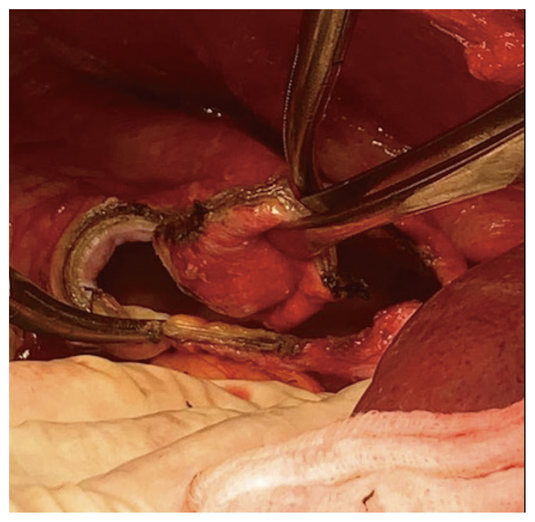
Type 3 liver mobilisation and full-thickness diaphragmatic resection with primary closure are the primary debulking surgical procedures used to manage advanced ovarian malignancies [1]. This requires a precise understanding of the anatomical landmarks, in addition to the surgical approach. Subspecialists must develop skills in this technique whilst avoiding potential complications, including injury to surrounding structures, nerves, and vessels, as well as post-operative morbidity. This video demonstrates the procedure in a 49-year-old female patient with a medical history of borderline serous ovarian cystadenoma. Investigation of symptoms revealed a CA125 level of 128, while imaging revealed a 9.8 cm complex ovarian mass with ascites, pelvic lymphadenopathy, possible metastatic liver serosa, and peritoneal nodularity. After multi-disciplinary team meeting and diagnostic laparoscopy, the patient underwent primary debulking of a suspected stage 3C ovarian malignancy (Table 1). During the laparotomy, extensive pelvic and peritoneal disease involving the right hemidiaphragm, bladder surface, appendix, and rectosigmoid were observed. On palpation, a diaphragmatic nodule raised the suspicion of full-thickness involvement with no evident cleavage plane in consideration of diaphragmatic stripping. Following total abdominal hysterectomy, salpingo-oophorectomy, omentectomy, appendectomy, and pelvic and para-aortic lymph node assessments (with pelvic lymph node dissection), type three liver mobilisation was performed using the Pringle maneouvre (Video 1, Table 2) [2,3]. Following the identification of the suprahepatic inferior vena cava and insertion of the right hepatic vein, the falciform and coronary ligaments were divided, allowing exposure of the right adrenal gland and retrohepatic inferior vena cava. The diaphragm was stripped and the muscle was opened to achieve full-thickness diaphragmatic resection with pleurectomy under direct vision of the surrounding lung parenchyma using an advanced energy device (Fig. 1) [4]. Primary closure was achieved using a single size 1 monofilament locking suture [5]. Diaphragmatic integrity was ensured by performing an air test and the Valsalva maneouvre. Further peritonectomy was performed on the bladder surface, rectosigmoid, and Morrison’s pouch, and a suspected port-site nodule (following diagnostic laparoscopy) was excised, achieving zero residual macroscopic disease. Final histology confirmed a serous borderline tumour with invasive implants within the port site nodule (stage 4A), warranting a medical oncology referral. Here, we highlight a challenging case requiring advanced surgical skills and knowledge, with specific consideration of intraoperative multidisciplinary decision-making regarding operative morbidity and mortality. The procedure was performed after consultation with the hepatobiliary and colorectal teams to achieve optimal cytoreduction and R0.

Notes

Conflict of interest

No conflict of interest declared.

Ethical approval

Not required.

Patient consent

Informed consent has been obtained.

Funding information

None.

Video clip

Video can be found with this article online at https://doi.org/10.5468/ogs.23029.

Fig. 1
Advanced energy device excising full thickness diaphragmatic nodules to include pleurectomy, ensuring appropriate macroscopic excision margins.
ogs-23029f1.jpg
Table 1
Procedure materials
Positioning
 Patient is placed in modified Lloyd Davis position with gluteal fold at the end of the table to allow access to perineum
Draping
 Abdominal skin preparation from mid chest to mid thighs
 Under buttocks, legs, sides, top and bottom
 Vaginal preparation & Foley’s catheter insertion
Instruments
 Bookwalter retractor system
 Monopolar pencil on extension set to 35 units cut and coagulation
 Bipolar scissors energy set to 60 units
 Ligasure impact advanced energy device
 Russian tissue and Robert artery forceps
 Number 1 Maxon suture
Table 2
Procedure summary
Ensure adequate access to the upper abdomen
Assessment of anatomy
 Identification of suprahepatic IVC
 Identification of right hepatic vein insertion
 Division of falciform and coronary ligaments
 Exposure of right adrenal gland and retrohepatic IVC
Type three liver mobilisation
 Pringle manoeuvre
Diaphragmatic resection
 Peritoneal stripping
 Muscle layer opened
 Full thickness resection using advanced energy device
 Direct vision lung parenchyma
Primary closure
 Number 1 locking Maxon suture
 Integrity assessment using air test and valsalva manoeuvre

IVC, inferior vena cava.

References

1. Buruiana FE, Ismail L, Ferrari F, Majd HS. The role of ultra-radical surgery in the management of advanced ovarian cancer: state of the art [Internet] London: IntechOpen; c2021 [cited 2022 Aug 3]. Available from: https://www.intechopen.com/chapters/78061 .

2. Tozzi R, Traill Z, Garruto Campanile R, Ferrari F, Soleymani Majd H, Nieuwstad J, et al. Porta hepatis peritonectomy and hepato-celiac lymphadenectomy in patients with stage IIIC-IV ovarian cancer: diagnostic pathway, surgical technique and outcomes. Gynecol Oncol 2016;143:35-9.
crossref pmid
3. Harter P, Sehouli J, Lorusso D, Reuss A, Vergote I, Marth C, et al. A randomized trial of lymphadenectomy in patients with advanced ovarian neoplasms. N Engl J Med 2019;380:822-32.
crossref pmid
4. Soleymani Majd H, Ferrari F, Manek S, Gubbala K, Campanile RG, Hardern K, et al. Diaphragmatic peritonectomy vs. full thickness resection with pleurectomy during visceral-peritoneal debulking (VPD) in 100 consecutive patients with stage IIIC-IV ovarian cancer: a surgical-histological analysis. Gynecol Oncol 2016;140:430-5.
crossref pmid
5. Addley S, Yao DS, Soleymani Majd H. Primary diaphragmatic closure following diaphragmatic resection and cardiophrenic lymph node dissection during interval debulking surgery for advanced ovarian malignancy. Gynecol Oncol Rep 2021;36:100744.
crossref pmid pmc
TOOLS
Share :
Facebook Twitter Linked In Google+ Line it
METRICS Graph View
  • 0 Crossref
  •     Scopus
  • 1,661 View
  • 96 Download
Related articles in Obstet Gynecol Sci


ABOUT
ARTICLE & TOPICS
Article category

Browse all articles >

Topics

Browse all articles >

BROWSE ARTICLES
POLICY
FOR CONTRIBUTORS
Editorial Office
4th Floor, 36 Gangnam-daero 132-gil, Gangnam-gu, Seoul 06044, Korea.
Tel: +82-2-2266-7238    Fax: +82-2-3445-2440    E-mail: journal@ogscience.org                

Copyright © 2024 by Korean Society of Obstetrics and Gynecology.

Developed in M2PI

Close layer
prev next