Evaluation of maternal rhesus blood type as a risk factor in adverse pregnancy outcomes in Korea: a nationwide health insurance database study
Article information
Abstract
Objectives
The current study aimed to investigate whether pregnancy outcomes are affected by maternal rhesus (Rh) status by comparing the primigravida pregnancy outcomes of Rh-negative women with those of Rh-positive women.
Methods
The study data were collected from the Korea National Health Insurance Claims Database and the National Health Screening Program for Infants and Children. In total, 1,664,882 primigravida women who gave birth between January 1, 2007 and December 31, 2014, were enrolled in this study. As the risk and severity of sensitization response increases with each subsequent pregnancy, only primigravida women were enrolled. The patients were divided into 2 groups according to Rh status, and the pregnancy outcomes were compared.
Results
In total, 1,661,320 women in the Rh-positive group and 3,290 in the Rh-negative group were assessed. With regard to adverse pregnancy outcomes, there was no statistically significant difference between the 2 groups in terms of the prevalence of preeclampsia, postpartum hemorrhage, abruptio placenta, placenta previa, and uterine artery embolization. A univariate analysis revealed that none of the adverse pregnancy outcomes were significantly correlated to Rh status (preeclampsia: odds ratio [OR], 1.00, 95% confidence interval [CI], 0.81–1.23; postpartum hemorrhage: OR, 1.10, 95% CI, 0.98–1.24; abruptio placenta: OR, 0.80, 95% CI, 0.46–1.37; and placenta previa: OR, 1.08, 95% CI, 0.78–1.42). The adjusted ORs of postpartum hemorrhage and preterm birth did not significantly differ.
Conclusion
Maternal Rh status is not associated with adverse outcomes in primigravida women.
Introduction
A person’s blood type is determined according to the specific antigen types on the erythrocyte membranes of red blood cells (RBCs). The 2 main factors that determine the blood type are ABO (A, B, AB, and O) and rhesus (Rh) (positive or negative). Karl Landsteiner first discovered the ABO blood group system in 1900 when he was investigating the causes of some fatal transfusions [1]. The ABO blood group system was named according to the different agglutinins, or blood group antigens, including A, B, and H (or O) antigens, on the surface of human RBCs [1]. Based on the presence or absence of the Rh factor, the blood group system is called the Rh blood group system. Rh represents the first 2 letters of the name Macacus Rhesus. In 1940, during animal experiments, Landsteiner and other scientists discovered that rhesus monkeys and most human RBCs have antigenic Rh blood types, and this was used in naming the system [2]. With continuous studies of the Rh blood groups, the Rh blood group system was found to be the most complex system in the RBCs [3]. The discovery of the Rh blood type has played an important role not only in guiding blood transfusions more scientifically but also in improving experimental diagnoses and clinical immunotherapy.
The blood type may affect human health and diseases with a wide range of expression in human cells and tissues, including platelets, epithelium, and vascular endothelium [4,5]. Therefore, several studies showed the clinical significance of the biological characteristics of the ABO blood system, particularly with regard to cancer, cardiovascular disease, and pregnancy-related disease [6-8]. Numerous reports revealed that the ABO blood group may be associated with some risk factors for unfavorable pregnancy outcomes [9].
However, data about whether maternal Rh blood type alone, without consideration of alloimmune sensitization, is associated with the development of pregnancy-related diseases are limited. Moreover, the Rh-negative population is extremely small. Thus, a large population must be evaluated to assess the role of Rh blood type in identifying women at risk of developing pregnancy-related complications.
Thus, this study aimed to investigate whether pregnancy outcomes are affected by maternal Rh status by comparing the primigravida pregnancy outcomes of Rh-negative women with those of Rh-positive women in a nationwide population study.
Materials and methods
1. Health care in Korea
Approximately 97% of the Korean population is enrolled in the Korea National Health Insurance (KNHI) program. All claims data are stored in the KNHI claims database. As part of the KNHI system, a National Health Screening Program for Infants and Children (NHSP-IC) was started in 2007, and it includes information about physical examination findings, anthropometric measurements, and developmental screening results after birth. This study used information from the KNHI claims database to identify all women who gave birth between January 1, 2007 and December 31, 2014. Moreover, whether these women were Rh-positive or Rh-negative based on the applicable codes from the International Classification of Disease, 10th Revision was assessed. The data of women who met the inclusion criteria were linked to those of their offspring in the NHSP-IC database.
2. Dataset and outcomes
Fig. 1 shows the flowchart of participant enrollment. Using the KNHI claims data, we identified all women who gave birth between January 1, 2007 and December 31, 2014 (n=3,383,282). The maternal datasets were merged with those in the NHSP-IC database. Multigravida women (n=1,650,500), those whose offspring had not undergone NHSP-IC health examinations (n=66,839), and those with missing data (n=1,061) were excluded from the study. Data regarding maternal and offspring outcomes were extracted. The information included pregnancy outcomes, such as parity, type of delivery, pulmonary embolism, postpartum hemorrhage, abruptio placenta, placenta previa, and uterine artery embolization. Furthermore, the characteristics of the neonates, such as sex, whether delivered preterm, and birth weight, were evaluated. Preterm birth was defined as birth at a gestational age <37 weeks, low birth weight (LBW) as birth weight <2.5 kg, and large for gestational age (LGA) as birthweight >4.0 kg.
3. Statistical analysis
Continuous and categorical variables were expressed as mean±standard deviation and number (percentage), respectively. The clinical characteristics of the participants were compared using t-test for continuous variables and the χ2 test for categorical variables. The risks of Rh-negative blood type were evaluated via multiple regression analyses. The risk of postpartum hemorrhage and preterm birth was adjusted for each related risk factor (postpartum hemorrhage: age, multiple pregnancy, cesarean section, preterm birth, LBW, LGA, preeclampsia, abruptio placenta, and placenta previa; preterm birth: age, multiple pregnancy, and preeclampsia). All tests were 2-sided, and a P-value <0.05 was considered statistically significant. Statistical analyses were performed using SAS for Windows (version 9.4; SAS, Cary, NC, USA).
Results
1. Baseline characteristics of the study population
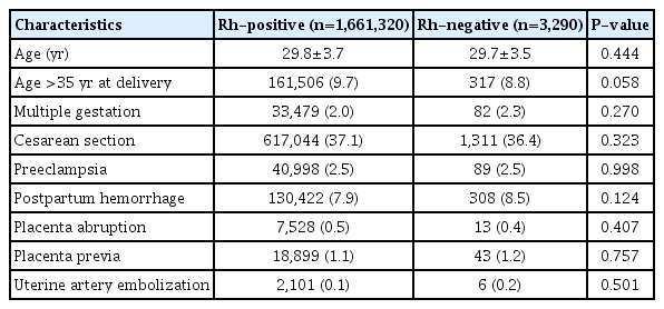
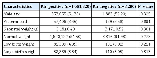
The clinical characteristics of the participants are shown in Table 1. There were no significant differences in terms of age at delivery and multiple pregnancy and cesarean section rates between the Rh-positive group (n=1,661,320) and Rh-negative group (n=3,290). Moreover, adverse delivery outcomes did not significantly differ between the 2 groups. Moreover, preterm birth rates and birth weight (corrected for gestational age) did not significantly differ between the Rh-positive and Rh-negative groups (Table 2).
2. Univariate regression analysis of Rh blood type as a risk factor for pregnancy outcomes
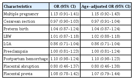
Univariate regression analyses were performed to evaluate the associations between Rh blood type and various clinical outcomes (Table 3). The Rh blood type was not a risk factor for preterm birth (odds ratio [OR], 1.13, 95% confidence interval [CI], 0.91–1.41), LBW (OR, 1.01, 95% CI, 0.87–1.18), and LGA (OR, 0.86, 95% CI, 0.71–1.04). Rh blood type was not a risk factor for adverse pregnancy outcomes (preeclampsia: OR, 1.00, 95% CI, 0.81–1.23; postpartum hemorrhage: OR, 1.10, 95% CI, 0.98–1.24; abruptio placenta: OR, 0.80, 95% CI, 0.46–1.37; and placenta previa: OR, 1.08, 95% CI, 0.78–1.42). Moreover, age-adjusted univariate regression analyses revealed no significant relationships between the Rh blood type and these factors.
3. Multiple regression analysis of rhesus blood type as a risk factor for postpartum hemorrhage and preterm birth
Based on the univariate analysis, the adjusted ORs for postpartum hemorrhage and preterm birth were analyzed because there was no significant relationship between Rh blood type and clinical outcomes (Table 4). The risk factors for postpartum hemorrhage were age, multiple pregnancy, cesarean section, preterm birth, LBW, LGA, preeclampsia, abruptio placenta, and placenta previa. Preterm birth was adjusted for age, multiple pregnancy, and preeclampsia. After adjusting for the factors, no relationships were found between Rh blood type and postpartum hemorrhage (OR, 1.10, 95% CI, 0.98–1.23) and preterm birth (OR, 0.02, 95% CI, 0.85–1.23).
Discussion
The proportions of blood type differ according to race, region, and ethnicity. In general, Africa, the Middle East, Europe, India, and Central Asia have higher Rh-negative blood rates than other regions. In Korea, Rh-negative is considered a rare blood type according to the Korean Red Cross standards, with an incidence of 0.4%; however, pregnancy can lead to several complications among Rh-negative women [10]. Currently, a non-O blood group may affect hemostatic balance disturbances and lead to an increased risk of embolization compared to an O blood group [11]. By contrast, another study showed a weak relationship between ABO blood type and gestational hypertension [12,13]. However, whether Rh blood type (negative or positive) is correlated to pregnancy outcomes is not known. Therefore, this study focused on the association between Rh-positive and Rh-negative blood groups and maternal pregnancy outcomes in 1,664,882 primigravida women in a large population study. The clinical characteristics did not differ significantly between Rh-positive (n=1,661,320) and Rh-negative women (n=3,290).
Several studies showed that the Rh blood type plays an important role in neonatal alloimmune disorders. The immune system of a Rh-negative mother considers Rh-positive fetal cells as foreign substances. Then, the mother’s body produces antibodies against fetal blood cells. Antibodies, such as IgG1 and IgG3, cross back through the placenta to the baby [14,15]. Then, they destroy the baby’s circulating RBCs, and this eventually develops into complications, such as preterm delivery, LBW, hydrops fetalis, hyperbilirubinemia, anemia, and bilirubin-induced neurological dysfunction [16-20].
In this study, there was no significant association between Rh blood type and neonatal outcomes, including preterm birth, LBW, and LGA. As previously mentioned, as the risk and severity of sensitization response increases with each subsequent pregnancy, we only included primigravida women in this analysis to reduce selection bias. During the first pregnancy, the initial exposure to fetal RBCs results in the formation of IgM antibodies, and these do not cross the placental barrier. Thus, no differences were observed in the first pregnancies of Rh-negative women [21]. Based on our results, the relationship between fetal gross and Rh blood type without an alloimmune effect is weak.
The World Health Organization reports that the major contributors to maternal death or maternal mortality are postpartum hemorrhage and hypertensive disorders [22]. Various studies have investigated whether there is an association between maternal ABO blood type and maternal adverse pregnancy outcomes [8,12,23-25]. In brief, maternal ABO blood type was correlated to the risk of preeclampsia. However, its association with GDM, preterm delivery, LBW, and SGA remains controversial.
Compared to the ABO types, studies about Rh blood type are limited. Thus, this study evaluated the differences in the rate of cesarean section, preeclampsia, postpartum hemorrhage, abruptio placenta, placenta previa, and uterine artery embolization among pregnant women with different Rh blood types. However, there was no significant association between Rh blood type and pregnancy outcomes, even preeclampsia, in primiparous women. Lee et al. [24] showed that Rh D-positive mothers had a slightly increased risk of preeclampsia in a large cohort study in Sweden (OR, 1.07, 95% CI, 1.03–1.10). However, in this study, multiparous women were enrolled, and only the Rh D antigen was evaluated. In a previous research, more than 50 Rh antigens have been identified at the serological level encoding 2 genes (RHD and RHCE). Among these antigens, Rh D, C, c, E, and e were considered the most clinically significant, and Rh D has the strongest antigenicity [26]. In our study, different findings were obtained, which might be attributed to the fact that we did not assess the differences in these antigens.
However, we did not find any associations between Rh blood type and adverse pregnancy outcomes. This result might be explained partly by the limitations of our research. First, this study only included Rh blood type classifications. However, the differences between the specific Rh blood type or the ABO classification were not assessed because the ABO blood group is not considered a disease category in the KNHI program. Second, as described above, the exclusion of multigravida pregnant women might have influenced the relatively varying results of the rates of preeclampsia compared to other previous studies. Third, with regard to neonatal outcomes, attention is now shifting towards shortand long-term morbidity [27]; however, in this study, only neonatal weight was included, and more detailed data are required to evaluate neonatal outcomes. To the best of our knowledge, this is the first nationwide retrospective research about the association between Rh blood type and adverse pregnancy outcomes in Korea. Hence, further studies must be conducted for a more detailed evaluation of the Rh blood type among pregnant women.
In conclusion, there were no significant differences in the risk of adverse pregnancy outcomes between Rh-positive and Rh-negative primigravida women in Korea.
Acknowledgements
This research was supported by the Basic Science Research Program through the National Research Foundation of Korea (NRF) funded by the Ministry of Science, ICT & Future Planning (NRF-2017R1C1B2010487). This research was supported by a grant from the Korea Health Technology R&D Project through the Korea Health Industry Development Institute (KHIDI) funded by the Ministry of Health & Welfare, Republic of Korea (grant number: HI17C1713).
Notes
Conflict of interest
No potential conflict of interest relevant to this article was reported.
Ethical approval
The study protocol was approved by the institutional review board of Korea University Medical Center (IRB number: 2019GR0256).
Patient consent
This paper is a retrospective study and does not require written informed consent.


